Posts

Image
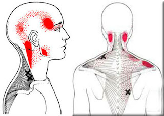
Cervicocephalic syndrome      Introduction Cervicocephalic syndrome (CCS) includes pain and stiffness of the upper cervical spine with associated headache. The syndrome frequently demonstrates symptoms of dizziness and often visual (e.g. nystagmus) or auditory disturbances (e.g. tinnitus). This term is outdated and not commonly used in clinical practice, although it may still be used in some parts of the world. Definition / Description These symptoms may be related to vertebrobasilar   insuffisiency where interference with the blood flow in the vertbral artery occurs when the neck is inclined to one side, rotated or extended. Clinically Relevant Anatomy The upper cervical complex consists of the atlanto - ocipital(jointC0-C1), the atlanto-axial (1joint-C2) and the superior aspect of C2. The vertebral arteries begin in the root of the neck. Usually the left artery is larger than the right. The cervical parts of the vertebral arteries ascen
Image
Glenohumeral The glenohumeral (GH) joint is a true synovial ball-and-socket style diarthroidal joint that is responsible for connecting the upper extremity to the trunk.  It is one of four joints that comprise the shoulder complex. This joint is formed from the combination of the humeral head and the glenoid fossa of the scapula. This joint is considered to be the most mobile and least stable joint in the body, and is the most commonly dislocated diarthoidal joint. Motions Available Abduction: elevation of the humerus on the glenoid in the frontal (coronal) plane. Flexion: forward and upward movement of the humerus on the glenoid in the sagittal plane. Extension: upward movemement of the humerus on the glenoid in the sagittal plane towards the rear of the body. Internal Rotation: rotation of the humerus on the glenoid in a medial direction. External Rotation: rotation of the humerus on the glenoid in a lateral direction. scapular plane abduction: elevation of the humerus o

Lumbar Vertebrae

Image
Lumbar Vertebrae Verebral Bodies   Lumbar Vertebrae Structure Lumbare The Lumbar Vertebrae are larger and heavier than vertebral bodies in other regions.  The lumbar vertebral body is kidney shaped when viewed superiorly, so is wider from side to side than from front to back, and a little thicker in front than in back with a thin cortial shell which surrounds cancellous bone. The posterior aspect of the vertebral body changes from slightly concave to slightly convex from L1 - L5 with an increasing diameter due to the increased load carried at each body.  The main weight of the body is carried by the vertebral bodies and disks. The lamina, facets and spinous process are major parts of the posterior elements that help guide the movement of the vertebrae and protect the spinal cord.  Vertebral Foramen The vertebral foramen is triangular in shape and is larger than in the thoracic vertebrae but smaller than in the cervical vertebrae.  Bony Structures   Pedicles  

Thoracic Vertebrae

Image
Thoracic Vertebrae The thoracic spine is part of the vertebral column that supports the chest area and provides posterior attachment for the ribs, some thoracic wall muscles, muscles of the upper limb abdomen and back. The thoraci spine lies between the cervical spine superiorly and the lumbar spine inferiorly. It consists of 12 thoracic vertebrae that are in some ways distinct from vertebrae found in other regions of the vertebral column Bodies The bodies of the thoracic vertebrae are medium sized and heart shaped. They are all weight bearing and generally increase in size from up downwards with an increase in the amount of weight that needs to be supported. The heads possess articular facets on their sides for articulation with the heads of the ribs Pedicles These project from the posterolateral surfaces of the bodies of the ribs and meet posteriorly with the laminae. The pedicles and laminae on either side form the neural arch which together with the posterior surfac